• Call: 022-22040711
  • Mail: info@cyreseye.com
  • Sun: Closed
  • Home
  • About
  • Services
    • Cataract
    • Glaucoma
    • Refractive – SMILE
    • Refractive – LASIK
    • Kerotoconus and C3R
    • Corneal Transplants DSAEK/DMEK/DALK
    • Vitreoretinal
    • Low Vision Clinic
    • Contact Lenses
    • Pediatric Dept Squint and Lazy Eye
  • Awards
  • Gallery
  • News
  • Testimonials
  • Ask a Question
  • Contact Us
Book an Appointment

Ask Us Form

Corneal Transplants DSAEK/DMEK/DALK

  • Home
  • Services
  • Corneal Transplants DSAEK/DMEK/DALK

Services

  • Cataract
  • Glaucoma
  • Refractive – SMILE
  • Refractive – LASIK
  • Corneal Transplants DSAEK/DMEK/DALK
  • Kerotoconus and C3R
  • Vitreoretinal
  • Low Vision Clinic
  • Kerotoconus and C3R
  • Contact Lenses
  • Pediatric Dept Squint and Lazy Eye
#
Corneal Transplants DSAEK/DMEK/DALK
Corneal Transplants -The Gift of Sight

The cornea is the clear front window of the eye. When it becomes hazy due to disease for eg Fuchs endothelial dystrophy or other corneal diseases or it gets injured by trauma or infection like a non-healing corneal ulcer set in the only option before us is to do a corneal transplant.

In the old days before 2000, the only option was a full-thickness corneal transplant called Penetrating Keratoplasty. Since then it was discovered that is the disease was in the front part of the eye we could remove only the front 4/5ths called DALK and if the disease was only at the back part of the cornea in the endothelial cells then we could.

1.Penetrating Keratoplasty-PK
What is a Corneal Transplant: Full Thickness

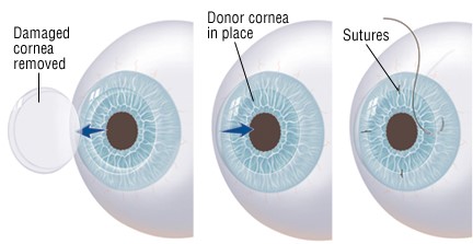
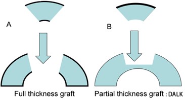
The full-thickness corneal transplant, or Penetrating Keratoplasty (PK), originated over 100 years ago. The procedure involves the transplantation of donor corneal tissue to replace the patient’s pre-existing, damaged central cornea. Usually, we remove the centra 7.5mm. This procedure is referred to as a full-thickness transplant because the entire damaged cornea is replaced by the donor cornea. There are a variety of conditions that can result in the need for a PK, including corneal swelling, scarring or distortion. These conditions blur the vision and may result from genetic conditions, infection or trauma.

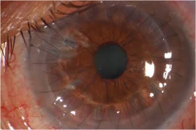
Corneal transplantation, when indicated, can help to restore functional vision in an eye with corneal damage. However, like all surgeries, there is a risk that must be balanced against the degree of difficulty the patient is experiencing as a result of his or her corneal disorder. The most common threat to the success of a corneal transplant is graft rejection. Rejection occurs in about 10% of cases, but if risk factors are present the rate of rejection may be much higher for some individuals. In most instances, episodes of rejection can be controlled by topical steroid eye drops or other forms of medicine; however, rejection may result in a permanent swelling of the donor cornea causing blurred vision, leading to the need for another full-thickness transplant. Sutures are used to hold the donor corneal tissue in place during the recovery period. Occasionally, the sutures can become loose, which may result in infection or induce astigmatism. It is impossible to predict the glasses prescription that will result after a corneal transplant. It is common for a significant amount of farsightedness, nearsightedness or astigmatism to be present following a full-thickness cornea transplant. In some cases, the glass prescription is strong enough that eyeglasses alone cannot offer adequate vision or balance with the other eye. For these individuals, contact lenses or additional surgery may help to bring the eyes into focus and balance.

Full-thickness transplants are vulnerable to a traumatic injury that may result in rupturing the eye (even years after the original transplant) because the transplant never heals as strong as a normal cornea. Therefore, extra caution and some restrictions are required for life after undergoing corneal transplantation, which must be considered prior to surgery. Typically, the visual recovery time following a full-thickness transplant is 2 years. Patients who undergo full-thickness corneal transplants may be on an eye drop regimen usually forever, following surgery.

Till a few years ago we were using a sharp curved metal blade called a “trephine” to cut the cornea.

Today we use a Femtosecond laser. This is called LACT, laser-assisted corneal transplant.

2. DALK-DEEP Anterior Lamellar Keratoplasty

Deep Anterior Lamellar Keratoplasty (DALK) can be a good option to consider when the innermost layers of the cornea are healthy, but the front most layers of the cornea are the problem. Common indications for this DALK_are primarily keratoconus and corneal scars. In DALK, the patient’s inner corneal layer (the endothelium) is left intact and all other layers of the cornea are removed. There are benefits with DALK: fewer postoperative complications, less incidence of rejection, and shorter use of topical steroid treatment.

A common threat to the success of a corneal transplant is graft rejection. Rejection occurs in about 10% of cases of traditional Penetrating Keratoplasty (PK). Rejection most commonly arises from the donor endothelium. The other layers of a transplanted cornea are much less likely to invite rejection. In DALK, a person’s endothelium is left intact and this lowers the incidence of rejection.

3.DSAEK-Descemet Stripping Automated Endothelial Keratoplasty
Descemet Membrane Endothelial Keratoplasty (DMEK)

DSAEK and DMEK are indicated when there is corneal clouding due to a dysfunction of the endothelium but where the other layers of the cornea remain healthy. With Fuchs’ endothelial dystrophy or when there is swelling of the cornea after cataract surgery, it is the endothelium that is compromised.
Fuchs dystrophy is very common in India especially in small communities like Parsees or Other small insular communities that intermarry.

What is the endothelium and how does it work?

The cornea is the clear dome that makes up the front part of the eye. It is about 1/10th of a centimeter thick and is composed of three primary layers: the epithelium, the stroma, and the endothelium. The epithelium is a thin surface layer. The stroma is composed mostly of fibrous proteins and acts like a sponge, absorbing fluid from inside the eye. The endothelium is a single layer of cells coating the inside portion of the cornea. Its job is to provide nutrients to the cells in the stroma and to make sure that the stroma has just the right amount of fluids. Endothelial cells pump fluid out of the cornea Too much fluid in the stroma can cause swelling of the cornea and decreased vision.

What is Endothelial Failure?

The cells of the corneal endothelium are very fragile. Two of the most common causes of endothelial failure are disease (like Fuchs’ Dystrophy) and trauma following cataract or glaucoma surgery. Once an endothelial cell dies it will not grow back. If an eye loses too many endothelial cells, it is not able to maintain the proper corneal thickness and clarity.

Replacement of the diseased endothelial layer in DSAEK surgery

Descemet’s Stripping Automated Endothelial Keratoplasty (DSAEK) is a partial thickness corneal transplant. During this procedure, the patient’s damaged endothelial layer is isolated and removed. A thin layer of donor corneal tissue that contains the healthy, endothelial layer is inserted onto the back surface of the patient’s cornea through a 3 mm tunnel An air bubble is then inserted into the front of the eye to hold the donor tissue in position. The air bubble is naturally absorbed by the eye within the first 24-48 hours following surgery. In most cases, the donor tissue remains in a good position as it heals in place but in some cases, the tissue can dislocate and a second air bubble may be needed to reposition the donor(known as rebubbling) or it may need to be replaced with new donor tissue. Typically, there are no sutures required to hold the new cornea in place.

This technique holds many advantages over the traditional full-thickness corneal transplant. Smaller surgical incisions and fewer sutures lead to faster recovery time. In fact, most patients begin to regain improved vision within the first few weeks or months following DSAEK compared with much longer recovery times after a full-thickness transplant. Typically, the glasses prescription after surgery only changes a modest amount (small plus number) by contrast with the relatively unpredictable amount of change associated with full-thickness corneal transplants. An eye that has undergone DSAEK is less vulnerable to rupturing (as compared to an eye that has undergone a full-thickness transplant) because the smaller operative wound is more stable, heals more completely and is less likely to break open due to trauma. Additionally, there appears to be a lower incidence of donor graft rejection because only a portion of the cornea is transplanted. Overall, for the appropriate candidate, DSAEK offers an excellent alternative to traditional corneal transplantation techniques with faster visual recovery and lower risk.

A new variant that cannot be done in some cases is called DMEK. Here the layer is even thinner than DSAEK and he visual recovery faster and better. However, it cannot be done in all cases. When it cannot be done then DSAEK is a better alternative.

DSAEK and DMEK are indicated when there is corneal clouding due to a dysfunction of the endothelium but where the other layers of the cornea remain healthy. With Fuchs’ endothelial dystrophy or when there is swelling of the cornea after cataract surgery, it is the endothelium that is compromised.

Dr Cyres K . Mehta’s International eye centre combines the skill and expertise of an internationally famous surgeon with the best technology and lasers that money can buy. Everything you see here is simply the best in the world. Nothing but the best for your eyes is our mission statement.

Services

  • Cataract
  • Glaucoma
  • Refractive – SMILE
  • Refractive – LASIK
  • Corneal Transplants DSAEK/DMEK/DALK
  • Kerotoconus and C3R
  • Vitreoretinal
  • Low Vision Clinic
  • Kerotoconus and C3R
  • Contact Lenses
  • Pediatric Dept Squint and Lazy Eye

Contact Details

202 , Ramnimi Building, Mandlik Road, Cotton World Building, Next To Indigo Restaurant, Colaba, Mumbai- 400001, India

  • Tell +91-22-22040711
  • Email info@cyreseye.com
  • Email cyresmehta@yahoo.com
  • Website cyreseye.com
Get Map Direction
Copyright © Dr. Cyres Mehta 2020. All Rights Reserved
Terms & Conditions | Privacy Policy