• Call: 022-22040711
  • Mail: info@cyreseye.com
  • Sun: Closed
  • Home
  • About
  • Services
    • Cataract
    • Glaucoma
    • Refractive – SMILE
    • Refractive – LASIK
    • Kerotoconus and C3R
    • Corneal Transplants DSAEK/DMEK/DALK
    • Vitreoretinal
    • Low Vision Clinic
    • Contact Lenses
    • Pediatric Dept Squint and Lazy Eye
  • Awards
  • Gallery
  • News
  • Testimonials
  • Ask a Question
  • Contact Us
Book an Appointment

Ask Us Form

Kerotoconus and C3R

  • Home
  • Services
  • Kerotoconus and C3R

Services

  • Cataract
  • Glaucoma
  • Refractive – SMILE
  • Refractive – LASIK
  • Corneal Transplants DSAEK/DMEK/DALK
  • Kerotoconus and C3R
  • Vitreoretinal
  • Low Vision Clinic
  • Kerotoconus and C3R
  • Contact Lenses
  • Pediatric Dept Squint and Lazy Eye
#
Kerotoconus and C3R

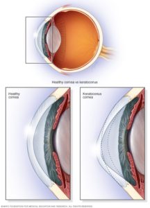
Keratoconus (KC) is a disorder of the eye which results in progressive thinning of the cornea over time. This can result in blurry vision, double vision, astigmatism, and light sensitivity. Usually both eyes are affected though one eye can be much more advanced than the other.

Keratoconus affects about 1 in 2,000 people. It occurs most commonly in late childhood to early adulthood. While it occurs in all populations it may be more frequent in certain ethnic groups such as those of Asian descent. The word is from the Greek kéras meaning cornea and the Latin cōnus meaning cone.

Eye rubbing and childhood eye allergies play a role in the development of keratoconus. The underlying mechanism involves changes of the cornea to a cone shape.

Keratoconus affects about 1 in 2,000 people. It occurs most commonly in late childhood to early adulthood. While it occurs in all populations it may be more frequent in certain ethnic groups such as those of Asian descent. The word is from the Greek kéras meaning cornea and the Latin cōnus meaning cone.

Signs and Symptoms

Simulation of the multiple images seen by a person with keratoconus.

People with early keratoconus typically notice a minor blurring of their vision and come to their clinician seeking corrective lenses for reading or driving. The vision can get progressively worse and the patient notices that even with frequent change glasses the vision isn’t improving. As the disease advances photophobia (sensitivity to bright light) and eye strain from squinting in order to read are common.

The classic symptom of keratoconus is the perception of multiple “ghost” images, known as monocular polyopia. The visual distortion experienced by the person comes from two sources, one being the irregular deformation of the surface of the cornea, and the other being scarring that occurs on it. These factors act to form regions on the cornea that map an image to different locations on the retina.

Genetics

Six genes have been found to be associated with the condition. These genes include BANP-ZNF469, COL4A4, FOXO1, FNDC3B, IMMP2L, and RXRA-COL5A1.

Pathophysiology

A genetic predisposition to keratoconus has been observed, with the disease running in certain families, and incidences reported of concordance in identical twins. Keratoconus is diagnosed more often in people with Down’s syndrome, though the reasons for this link have not yet been determined.

Keratoconus has been associated with atopic diseases, which include asthma, allergies, and eczema, and it is not uncommon for several or all of these diseases to affect one person. A number of studies suggest vigorous eye rubbing contributes to the progression of keratoconus, and people should be discouraged from the practice.

Diagnosis

A schematic diagram showing the change in the cornea with keratoconus.

A Corneal topographer, used for mapping the surface curvature of the cornea.

The eye doctor will perform a series of examinations and tests.

1. Slit-lamp examination
2. Topography
3. Corneal OCT


Stages

Corneal topography showing stage II keratoconus

Once keratoconus has been diagnosed, its degree may be classified by several metrics:

  • The steepness of greatest curvature from ‘mild’ (< 45D), ‘advanced’ (up to 52 D) or ‘severe’ (> 52 D);
  • The morphology of the cone: ‘nipple’ (small: 5 mm and near-central), ‘oval’ (larger, below-center and often sagging), or ‘globus’ (more than 75% of cornea affected);
  • The corneal thickness from mild (> 506 μm) to advanced (< 446 μm).
Management
General Treatment

For visual improvement and astigmatism management, spectacles or soft toric contact lenses in mild cases can be used. Rigid gas permeable contact lenses are needed in the majority of cases to neutralize the irregular corneal astigmatism. The majority of patients that can wear hard or gas-permeable contact lenses have a dramatic improvement in their vision. Specialty contact lenses have been developed to better fit the irregular and steep corneas found in keratoconus; these include ( but not limited to) Rosek, custom-designed contact lenses ( based on topography and/or wavefront measurements), semi-scleral contact lenses, piggyback lens use ( soft and hard lens used at the same time), and scleral lenses. Those patients that become contact lens intolerant or do not have an acceptable vision, typically from central scaring, can proceed to surgical alternatives.

Accelerated Corneal Cross Linking, C3R

The primary treatment for progressive keratoconus, or keratoconus in young patients likely to progress at some point, is corneal collagen cross-linking. This treatment, approved by the FDA in the Spring of 2016 and CE marked 10 years earlier throughout Europe in 2006, uses riboflavin and UV light to induce hardening of the otherwise weak keratoconic cornea. Traditional corneal collagen cross-linking involves a minimally invasive treatment through the removal of the corneal epithelium followed by soaking of the cornea in riboflavin drops and a slow exposure of the cornea to UV light. Variations of this conventional “epithelium-off” technique involve higher intensities of UV for shorter time periods (accelerated cross-linking).

What we are doing till July 22, was the the “Cretan Protocol”-This is using an excimer laser to ablate the epithelium first and then soak the cornea for 15 minutes and then do an accelerated CXL for 8 minutes. This also can decrease the cylinder of the person as we customize the ablation profile and this makes them see better.

NEW AVEDRO ROBOTIC SYSTEM -GOOD NEWS FOR KERATOCONUS PATIENTS.
PEOPLE WHO EARLIER COULD NOT UNDERGO LASIK OR SMILE NUMBER CORRECTION NOW CAN
BETTER VISION IN 90 SECONDS.

In July ’22 we have acquired the FIRST updated automated robotic AVEDRO KXL II cornea system in Maharashtra- What is it used for

Keratoconus the cornea or the clear front window of the eye due to genetic reasons is weak and thins out and bulges. When this happens the patient gets a huge number and bad vision which cannot be corrected with specs or conventional laser vision correction- (LASIK, CONTOURA OR SMILE) because the “bulge” isn’t regular and so now with the new AVEDRO system we can perform painless automated correction in only  90 seconds.
Some people have thin corneas where we cannot perform Lasik or SMILE to remove the number. With AVEDRO we can help many  of these people and do a Lasik XTRA or a SMILE XTRA in just 90 seconds . Here we will perform the laser vision correction procedure,put a special Riboflavin solution called VIBEX XTRA on the eye and then apply the AVEDRO laser for 90 seconds only.

Earlier old systems would put energy on the eye for 7-10 minutes in a continuous manner. Here some times the patients would develop haze and poor vision. This because when you apply continuous laser, Today AVEDRO system uses a pulsed energy approach. Here in between pulses oxygen goes into the cornea and refreshes the cells. So, no cell death or haze will occur and patient will have better vision. This takes only 90 seconds!.

OLD CROSS LINKING SYSTEMS

Most centers even today scrape the cornea first  with a brush or a blade ,which we never did ,in fact we always used a laser to  remove the top layer which is still a bit painful for 3 days .
Total treatment time I sometimes 20 minutes or more.
We have to go slow as energy is continuous, if we increase energy there is cell death.
We cannot combine safely with LASIK or SMILE.

AVEDRO FULLY AUTOMATED SYSTEM

No need to remove corneal epithelium in most cases-we use PHOTREXA epi on solution which enters the cornea through intact epithelium-painless
High Energy for 90 seconds only
Unique patented pulsed energy delivery
Can be combined with LASIK and SMILE to fix number in patients who earlier couldn’t remove spectacle number.

Naturally AVEDRO is a great boon for suffering   patients. AVEDO is the most modern corneal cross linking system in the world today.

Read what the AMERICAN ACADEMY OF OPHTHALMOLOGY SAYS ABOUT CROSS LINKING.

Game Changer

The FDA’s step to approve crosslinking is “a game changer,” said Bennie H. Jeng, MD, at the University of Maryland School of Medicine in Baltimore. CXL “is the first available modality that can halt the progres¬sion of keratoconus. If we’re able to reach patients at a stage where they have decent vision and freeze them in that position for the rest of their lives, that’s groundbreaking.”
Two European studies have already reported significant reductions in the number of corneal transplants for KC since the introduction of CXL there. Dr. Jeng said
“I believe CXL is one of the most important innovations in anterior segment surgery since the introduction of excimer lasers,” said Alaa M. ElDanasoury, MD, at Magrabi Eye and Ear Hospitals in Saudi Arabia.

ORIGINAL OLD PROTOCOL

The protocol. The standard CXL protocol, known as the Dresden protocol, involves removal of the central corneal epithelium to allow better diffusion of the riboflavin into the stroma.
After de-epithelialization, a 0.1% riboflavin solution is applied to the cornea—every 1 to 3 minutes for 30 minutes, in a process called inhibition—until the stroma is completely penetrated. The cornea is then irradiated for 30 minutes with 370 nm UVA (a maximal wavelength for absorption by riboflavin) at a power of 3 milliwatts (mW)/cm.


Evolving Techniques

Although the Dresden protocol is supported by the most re¬search and clinical experience, it has 2 major disadvantages, especially in children, for whom cooperation and compliance are major issues:

1. The duration of the procedure, which runs approximately 1 hour.
2. The removal of the epithe¬lium to allow the riboflavin to penetrate the deeper corneal layers. De-epithelialization results in postoperative pain and a higher risk of infection, melting, scarring, and haziness, Dr. Rapuano said more modern options which let to the development of AVEDRO KXL II are

Accelerated CXL(CRETAN PROTOCOL)

One option is to reduce exposure time by increasing the power density of the irradiation. Anterior 300 μm of the cornea can be cross-linked to stabilize KC by increasing the power density to 9 mW/cm2 and decreasing irradiation time to 10 minute.

Transepithelial CXL

Investigators also have searched for methods to keep the epithelium on, by using different compositions of riboflavin solution to try to penetrate the cornea with an intact epithelium. earlier this did not work. Now with PHOTREXA and AVEDRO KXL II we can.

Surgery

Surgical options can include – Deep Anterior Lamellar Keratoplasty(DALK), Penetrating Keratoplasty.

DALK is the modern option for treating keratoconus. It involves the replacement of the central anterior cornea, leaving the patient’s endothelium intact. The advantages are that the risk of endothelial graft rejection is eliminated, and there is less risk of traumatic rupture of the globe in the incision, since the endothelium and Descemet’s and some stroma are left intact, and faster visual rehabilitation.

Penetrating keratoplasty has a high success rate and is the standard surgical treatment with a long track record of safety and efficacy.

Dr Cyres K . Mehta’s International eye centre combines the skill and expertise of an internationally famous surgeon with the best technology and lasers that money can buy. Everything you see here is simply the best in the world. Nothing but the best for your eyes is our mission statement.

Services

  • Cataract
  • Glaucoma
  • Refractive – SMILE
  • Refractive – LASIK
  • Corneal Transplants DSAEK/DMEK/DALK
  • Kerotoconus and C3R
  • Vitreoretinal
  • Low Vision Clinic
  • Kerotoconus and C3R
  • Contact Lenses
  • Pediatric Dept Squint and Lazy Eye

Contact Details

202 , Ramnimi Building, Mandlik Road, Cotton World Building, Next To Indigo Restaurant, Colaba, Mumbai- 400001, India

  • Tell +91-22-22040711
  • Email info@cyreseye.com
  • Email cyresmehta@yahoo.com
  • Website cyreseye.com
Get Map Direction
Copyright © Dr. Cyres Mehta 2020. All Rights Reserved
Terms & Conditions | Privacy Policy