• Call: 022-22040711
  • Mail: info@cyreseye.com
  • Sun: Closed
  • Home
  • About
  • Services
    • Cataract
    • Glaucoma
    • Refractive – SMILE
    • Refractive – LASIK
    • Kerotoconus and C3R
    • Corneal Transplants DSAEK/DMEK/DALK
    • Vitreoretinal
    • Low Vision Clinic
    • Contact Lenses
    • Pediatric Dept Squint and Lazy Eye
  • Awards
  • Gallery
  • News
  • Testimonials
  • Ask a Question
  • Contact Us
Book an Appointment

Ask Us Form

Vitreoretinal

  • Home
  • Services
  • Vitreoretinal

Services

  • Cataract
  • Glaucoma
  • Refractive – SMILE
  • Refractive – LASIK
  • Corneal Transplants DSAEK/DMEK/DALK
  • Kerotoconus and C3R
  • Vitreoretinal
  • Low Vision Clinic
  • Kerotoconus and C3R
  • Contact Lenses
  • Pediatric Dept Squint and Lazy Eye
#
Vitreoretinal

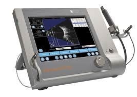
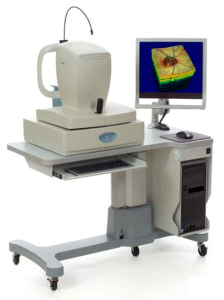
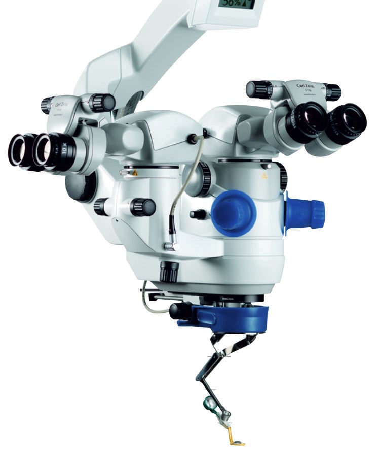
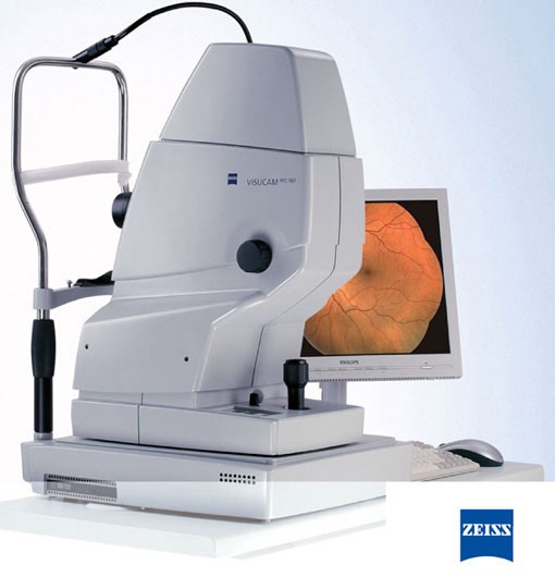
Dr. Cyres K Mehta’s International Eye Centre has made the biggest investment for our retina patients with the purchase of Zeiss Visucam 500 fundus camera,   the best and most advanced non-mydriatic fundus camera for taking pictures of the retina

This camera has a few advantages

  • You don’t have to dilate to take pictures of the retina
  • It will do  conventional Fluorescein angiography
  • Zeiss optics are the best in the world
  • It has Fundus autofluorescence(FAF) which can see the retina in great detail WITHOUT having to inject a dye into the patient. It can delineate membranes of macular degeneration and CSR lesions and is a great tool for noninvasive retina diagnostics
  • It has MPOD which is only present in this device (Macular pigment density). Using MPOD we can actually track the progress of fry macular degeneration which was hitherto impossible.

The Zeiss Visucam 500 fundus camera helps in the detection and management of sight-threatening diseases like age-related macular degeneration, diabetic retinopathy, glaucoma, hypertensive retinopathy, cancer of the eye and other retinal diseases.

Quantel Medical Compact Touch B-Scan

The COMPACT TOUCH combines the best image and measurement performance in its category with outstanding ergonomics. A possible 3 in 1 unit, It can meet just about all your Ultrasound needs.

  • Image of the globe and orbit (10 MHz B-scan)
  • Biometry
  • Pachymetry

Compact Touch is the best ultrasound b scan unit due to its exceptional image quality, its compact, lightweight ergonomics, and intuitive use.

The intuitive interface and touch-screen combined with compact size and high-level performance make this the benchmark product in terms of profitability and reliability.

High-quality b-scan images supporting your diagnosis. The quality of the image makes it possible to distinguish globe and orbit structures and to perform a detailed diagnosis of several different pathologies: cataract, the opacity of the vitreous, retinal detachment, etc.


The Avanti Optovue Angio OCT- First in Mumbai

Avanti Widefield OCT gives you new information on structures outside the traditional 6×6 mm cube, separates the retina into distinct layers for detailed assessment, offers views of the vitreous and deep choroid, and gives you the ability to monitor change over time. With the extensive information delivered by the Avanti System, you can tailor your approach to treatment and truly personalize patient care. I have acquired this first angio system in Mumbai. With a regular OCT we can only see the thickness of various layers of the retina Here with this system we can actually do angiography of the retina and actually see a membrane responsible for Wet macular degeneration which is amazing.

Membrane of Wet Macular Degeneration Seen Progressing:

Barrage Laser

The Barrage Laser is a GREEN  Laser treatment which is performed to strengthen weakened areas of the retina. Barrage laser treatment is also performed to seal retinal tears. Barrage laser treatment is an out-patient procedure where an intense beam of laser light is focused onto the retina.

Vitra Green Laser from France


Age Related Macular Degeneration (ARMD)

The center of the retina is called the “Macula”. When we read a book, or look at a vision chart or gaze at anything, we are using the macula to look at it. The rest of the retina is for peripheral vision. For example, when we drive down the street and are looking at the car in front of us, it is the macula that enables us to read the license plate of the car in front, but the cars on the sides are seen by the peripheral retina.

The macula is affected by a disease like any other part of the body. and most importantly it is affected by Age-Related Macular Degeneration.

Typically the elderly lady or gentleman has had their cataracts out a few years ago. While reading a novel, watching TV, or looking at the faces of his or her grown children realizes that all is not right. There is a grayish haze in the center of the vision that refuses to go away and seems to be getting worse as the month pass.

On reaching the eye doctor who performs a retinal exam, the patient is confronted with the diagnosis of ARMD.

Who is general is affected by ARMD

ARMD is quite a public health problem today. Many small communities like Parsis and Jews are overly affected by this due to their small genetic pool as well as our propensity to live long.

Everyone knows someone a friend, a spouse or a parent with this condition

In general, it’s been found that,

  • 20% of people age 65-74.
  • 40% of people age over 75 have macular degeneration.
  • Age-Related Macular Degeneration is a true public health issue even in the west, 14 million had Macular Degeneration in 1996 in the USA alone.
What question should I ask the doctor about ARMD
  • Ask the doctor if it is wet or dry. In Dry macular degeneration yellow spots called drusen appear in the macula. Ten to 15% of these patients progress to wet type. The dry type is slower progressing in general and does not lead to a sudden loss of vision.
  • In the wet type, a collection of blood vessels leaks fluid and blood into the retina. This reduces vision severely. In the wet type, bleeds can occur suddenly and the patient can swing from good to bad vision very quickly, sometimes in as little as a day.
What tests have to be done for ARMD

1.Photography of the Reina
2.Fundus Autofluorescence
3.Angio OCT

The total vision examination is performed. Next, the doctor will look at the retina with various lenses. He may take a photograph with a retina camera to document the current state of the disease.

The doctor will advise you 2 tests which will analyze the retina. Fluorescein Angiography was used to detect leaks. Nowadays instead we use a FAF on the Zeiss system and an Angio OCT test which will determine swelling and thickening of the retina presence of the membrane and leakage. These tests are simple but give a lot of information on the disease.

What's the treatment for ARMD

The dry type has no treatment! Usually, a powerful vitamin tablet with minerals is prescribed. Different formulations have been shown to have some use in arresting the progress of the disease.

Basically treatment is for the wet type.

  • Drugs called ANTI VEGF,s (AVASTIN / LUCENTIS /RAZUMAB/EYELET)are injected into the eye which makes the membrane shrink. Many patients have had useful vision improvement with this medicine. Sometimes this medicine is combined with PDT for better results.
  • A new telescope implant is implanted in the eye with a procedure similar to cataract surgery. In cases usually dry type, this has yielded good results in practice.
  • The new microchip has been invented which when implanted into the retina will amplify the vision.

Only your eye doctor can judge in your case which mode is appropriate.

What's the prevention of this disease entail ?

Studies performed in the UK showed that if the diet was rich in vitamins like Vitamin A, C, K, etc and trace elements like zinc and selenium, the persons had 25 % less chance of developing ARMD, So regularly take a good vitamin and mineral supplement. It’s a good idea to from middle age itself has a diet rich in red, yellow and green vegetables as they contain special compounds which may prevent the disease.

UV light and sun exposure is a major cause so please wear dark glasses in the sun as a rule.

Diabetes and Its Effect on the Eye

Introduction:

Diabetes is a disease characterized by high blood sugar levels. If you have diabetes, regular visits to your ophthalmologist for eye exams are important to avoid diabetes eye problems. High blood glucose increases the risk of diabetes eye problems. In fact, diabetes is the leading cause of blindness in adults age 20 to 74.

If you have eye problems and diabetes, don’t buy a new pair of glasses when you notice you have blurred vision. It could just be a temporary eye problem that develops rapidly with diabetes and is caused by high blood glucose levels.
High blood glucose in diabetes causes the lens of the eye to swell, which changes your ability to see. To correct this kind of eye problem, you need to get your blood glucose back into the target range (90-130 mg/dL before meals, and less than 180 mg/dL one to two hours after a meal). It may take as long as three months after your blood glucose is well controlled for your vision to fully get back to normal.

Blurred vision can also be a symptom of more serious eye problems with diabetes. The three major eye problems that people with diabetes may develop and should be aware of are cataracts, glaucoma, and retinopathy.

The eye is the only place in the body that by simply looking in with a scope we can judge the amount of diabetes in the body and even estimate kidney function due to diabetes and its disease.

What is Diabetic Cataract

The human lens in diabetes swells up as water enters it. The diabetic cataract is easily recognized by the eye doctor.

The treatment is simple. Through robotic laser lens replacement, the cataract is liquefied with a laser and sucked out. In its place, a special foldable lens is injected into the eye. In the old days, the eye took months to heal after cataract surgery in a diabetic as the cornea is weak in diabetics. Today with the new laser system, the recovery is much faster.

Can Glaucoma occur due to diabetes

Pressure in the eye goes up in diabetes as the lens swells up and occupies more space in the eye. Also, retinal damage causes new blood vessels to grow that choke up the outflow pathway of the eye so that fluid cannot flow out of the eye. Removing the cataract and or treating the retina with laser usually solves the problem.

Can Squint arise due to diabetes

In some cases when the blood sugar level goes very high the patient starts to see double as the nerves to the eye muscles get affected and the eye squints. The treatment of this is the treatment of the high blood sugar after which the squint slowly resolves itself.

Who gets diabetic retinopathy?

Anyone who has diabetes has a risk of acquiring diabetic retinopathy. Sadly, around 80 percent of all diabetics who have been diagnosed with diabetes for at least ten years will be a victim of the disorder. If diabetes is well controlled the retina may be normal even after 15 years and if it’s been poorly controlled even 10 years leads to significant visual loss.

What are the symptoms of diabetic retinopathy ?

Basically three stages.

  • Background stage- few dot and blot hemorrhages and microaneurysms, needs no treatment.
  • Pre proliferative stage -This is the stage where we must start laser treatment and or ANTI VEGF injections.
  • Proliferative stage-Treat urgently with injections and laser and sometimes vitrectomy surgery.

Exudative maculopathy and Hard exudates (swelling at the center of the retina) can occur at any stage and seriously impact vision. Maculopathy requires a laser and repeated injections.

Diabetic retinopathy can develop over a long period of time without any significant indicators. Although there are changes in retinal blood vessels, these may or may not be visible to an untrained eye. Essentially, vision will become blurred or lost completely when diabetic retinopathy is in full effect. The onset of this disorder can be slow, but it may impact a patient’s vision seemingly immediately. During the non-proliferative stage of diabetic retinopathy, aneurysms and leakages occur in the vessels producing swelling and leakage within the retina. This can result in blurry sight quality. These eye hemorrhages (bleeds) often occur multiple times and can take years to clear, if ever.

During proliferative diabetic retinopathy, the leakage described above has produced scarring and build up in the retina, often causing floaters along with a blurred vision. In aggravated cases of advanced diabetic retinopathy, a person may only be able to distinguish the contrast between dark and light in the affected eye(s). The new vessel growth, due to a lack of oxygen to the retina, is an unhealthy by-product of the abnormal blood vessel activity, and these vessels can begin to grow on the vitreous. Scar tissue growth can also be a development in this stage of diabetic retinopathy, which may create tearing and pulling on the retina. In this case, blindness is a very real concern.

What are the tests normally carried out for for Diabetic Retinopathy

Fluorescein Angiography was traditionally done where 3cc of a dye is injected into a vein on the hand. Photos of the retina are now taken with a special wavelength of light which makes the dye in the retinal vessel fluoresce and leakage is instantly seen and captured by a digital camera.

In some people, there is a little bit of nausea with the dye injection which soon passes. The urine turns brown as the dye exits the body over the next 2 days. So the patient should not worry if the urine looks “bloody”.

Patients with very advanced diabetes have bad kidney function. Since the dye leaves the body through the kidney it’s advisable to inform the eye doctor of the kidney changes so he can have a word with your diabetologist/physician or nephrologists.

The latest innovation Angio-OCT-Ocular Coherence Tomography

Here is just a minute, the retina can be analyzed without even touching the eye just by this system which uses a red laser to scan the eye retina and detect swelling and leakage. It’s very quick. A laser scans the central retina and reconstructs a picture of the different retinal layers as it bounces off them. It gives us information about leakage and swelling. In these systems, the laser scanner is so fast that it can track the movement of red blood cells through the capillaries. We have nearly stopped doing a traditional angiography because we don’t need to inject dye into the vein so we can avoid side effects of the die like fainting, vomiting, and kidney damage.

Treatment
1. Green / Yellow Laser Photocoagulation is still the mainstay of diabetic treatment.

How does the laser work? When we apply the laser to the eye it destroys part of the retina which produces a chemical called VEGF which is responsible for most of the bad effects of diabetes like leakage, swelling, and bleeding. This (VEGF) chemical produced by the retina makes the retina produce tiny defective blood vessels that keep leaking and bleeding. Also in diabetes, the blood flow in the capillaries of the retina is affected, so the retina does not get enough oxygen. Because it does not get enough oxygen it further releases more VEGF which grows more new defective blood vessels in an effort to better the oxygenation of the retina. So laser is applied to the peripheral retina. The laser causes localized scarring and that part of the retina is non-living and does not require oxygen. As it does not need more oxygen it hence stops producing VEGF and the new blood vessels regress.

Also, the laser is applied to seal leaking defective vessels that are already present.

Naturally, since laser has been applied to a large area of the retina the side vision decreases, and night vision decreases. Thus the central vision is preserved for a long time while side vision decreases. This is the compromise that has to be made in order to retain useful central vision in longstanding severe diabetic retinopathy.

2. Anti-VEGF drugs (Lucentis & Eyelea)

A new treatment is available whereby special drugs called ANTI-VEGF drugs (brand names like Lucentis, Eylea) are injected painlessly via a very fine needle as thin as a hair into the eye. This makes the new blood vessels which leak to subside. It’s an anti-VEGF drug. In Europe and the USA nowadays these medicines are injected monthly instead of applying too much laser. The advantage of medicine based treatment over just laser is that it preserves the side vision which laser destroys.
More details can be sought at drcr.net

3. Vitreo-Retinal Surgery

Once the retinopathy is going to enter the proliferative stage the best option is a surgery where the jelly at the back of the eye called vitreous is removed through a tiny opening and laser applied from inside the eye.

If there is persistent bleeding in the vitreous jelly,3 port pars plana vitrectomy is the best bet.

If diabetes has caused a retinal detachment, surgery to repair it is mandatory.

Surgical Equipment for Retina Surgery with International Eye Centre
1.Optikon Revolution-First in India

This is a very advanced expensive retina surgery machine. This machine can actually monitor in real-time the pressure gradient between the patients’ blood pressure and eye pressure. This is not available in any other system Cyres has visited Optikon in Rome and seen first hand the research center where the machine was developed and spoken and interacted with the scientists.

2. Zeiss Visalis 500 Retina Platform-First in India

This superlative system channels Zeiss 100 years of supremacy in eye care to offer unparalleled technology for retina surgery.

Retinal Viewing System-Zeiss Resight 700-First in India

Retina Detachment – A Brief Overview

  • A retinal detachment is a separation of the retina from its attachments to the underlying tissue within the eye.
  • Most retinal detachments are a result of a retinal break, hole, or tear. Most retinal breaks, holes, or tears are not caused by trauma (injury) but are due to preexisting factors such as high levels of myopia (nearsightedness), prior ocular surgery, and other eye diseases.
  • Flashing lights and floaters (tiny black dots moving around)may be the initial symptoms of a retinal detachment or of a retinal tear that precedes the detachment itself.
  • Early diagnosis and repair of retinal detachments are important since visual improvement is much greater when the retina is repaired before the macula or central area of the retina is detached.
  • The surgical repair of a retinal detachment is usually successful in reattaching the retina.
Rhegmatogenous Retinal Detachment

Most retinal detachments are a result of a retinal break, hole, or tear. A retinal detachment of this type is known as a rhegmatogenous retinal detachment. Most retinal breaks, holes, or tears are not a result of the injury. The majority of retinal breaks, holes, or tears are spontaneous, result when the vitreous gel pulls loose or separates from its attachment to the retina, usually in the peripheral parts of the retina. The vitreous is a clear gel that fills the rear two-thirds of the eye and occupies the space in front of the retina. As the vitreous gel pulls loose, it will sometimes exert pulling forces, known as traction, on the retina, and if the retina is weak, the retina will tear. Retinal tears are sometimes accompanied by bleeding if a retinal blood vessel is included in the tear. Everyone develops some shrinkage of the vitreous as they age, and many people develop separation of the vitreous from the retina as they get older. However, only a small percentage of these vitreous separations result in retinal tears.

Once the retina has torn, liquid from the vitreous gel can then pass through the tear and accumulate behind the retina. The buildup of fluid behind the retina is what separates (detaches) the retina from underlying layers in the back of the eye. As more of the liquid vitreous collects behind the retina, the extent of the retinal detachment can increase and involve the entire retina, leading to a total retinal detachment. A retinal detachment almost always affects only one eye at a time. The second eye, however, must be checked thoroughly for any signs of predisposing factors or existing retinal tears or holes that may lead to a retinal detachment in the future.

What are Retinal Detachment symptoms and signs?

Flashing lights (photopsia) and floaters (floating spots in the vision) may be the initial symptoms of a retinal detachment or of a retinal tear that precedes the detachment itself. Anyone who is beginning to experience these symptoms should see an eye doctor (ophthalmologist) for a retinal examination. In this exam, drops are used to dilate the patient’s pupils to make a more detailed exam easier. The symptoms of flashing lights and floaters may often be unassociated with a tear or detachment and can merely result from a separation of the vitreous gel from the retina. This condition is called a posterior vitreous detachment (PVD). Although a PVD occurs commonly, in the majority of cases, there are no tears associated with the condition.

The flashing lights are caused by the vitreous gel pulling on the retina or looseness of the vitreous, which allows the vitreous gel to bump against the retina. The lights are often described as resembling brief lightning streaks in the outside edges (periphery) of the eye. The floaters are caused by condensations (small solidifications) in the vitreous gel and frequently are described by patients as spots, strands, or little flies. Some people even want to use a flyswatter to eliminate these pesky floaters. There is no safe treatment to make the floaters disappear. Most people with complaints of floaters do not have tears of the retina or a retinal detachment.

If the patient experiences a shadow or curtain that affects any part of the vision of either eye, this can indicate that a retinal tear has progressed to a detached retina. In this situation, one should immediately consult an eye doctor since time can be critical. The goal for the ophthalmologist is to promptly make the diagnosis and treat the retinal tear or detachment before the central macular area of the retina detaches.

What are retinal detachment causes and risk factors?

Studies have shown that the incidence of retinal detachments caused by tears in the retina is fairly low, affecting approximately one in 10,000 people each year. Many retinal tears do not progress to retinal detachment. Nevertheless, many risk factors for developing retinal detachments are recognized, including certain diseases of the eyes (discussed below), complicated cataract surgery, and trauma to the eye. Retinal detachments can occur at any age but are most common in adults 40 years old and older who are highly myopic (high minus number) and also in older people who have undergone cataract surgery.

Which diseases of the eyes predispose to the development of a retinal detachment?
  • Lattice degeneration of the retina is a type of thinning of the outside edges of the retina, which occurs in 7%-10% of the general population. The lattice degeneration, so-called because the thinned retina resembles the crisscross pattern of a lattice, often contains small holes. Lattice degeneration is more common in people with nearsightedness (myopia). This tendency to lattice degeneration occurs because myopic eyes are larger than normal eyes and, therefore, the peripheral retina is stretched more thinly. Fortunately, only about 1% of patients with lattice degeneration go on to develop a retinal detachment. All patients with lattice degeneration of the retina should be informed about this condition and cautioned about the importance of seeing an ophthalmologist immediately if they develop sudden floaters, flashes, or a cloud in their vision.
  • High myopia (greater than 5 or 6 diopters of nearsightedness) increases the risk of a retinal detachment. In fact, the risk increases to 2.4% as compared to a 0.06% risk for a normal eye at 60 years of age. (Diopters are units of measurement that indicate the power of the lens to focus rays of light.) Complicated cataract surgery or other operations of the eye can further increase this risk in those with high myopia.
What other factors are associated with a retinal detachment?
  • Blunt trauma, as from a tennis ball or fist, or a penetrating injury by a sharp object to the eye can lead to a retinal detachment.
  • A family history of a detached retina that is non-traumatic in nature seems to indicate a genetic (inherited) tendency for developing retinal detachments.
  • Blunt trauma, as from a tennis ball or fist, or a penetrating injury by a sharp object to the eye can lead to a retinal detachment.
  • Diabetes that has been complicated by the development of proliferative diabetic retinopathy can lead to a type of retinal detachment that is caused by pulling on the retina (traction) alone, without a tear. Because of abnormal blood vessels and scar tissue on the retinal surface in some people with diabetic proliferative retinopathy, the retina can be lifted off (detached) from the back of the eye. In addition, the blood vessels may bleed into the vitreous gel. This detachment may involve either the peripheral or central area of the retina.
  • Non-rhegmatogenous retinal detachments are those that are not a result of a retinal break, hole, or tear. These include conditions known as exudative retinal detachments and tractional retinal detachments.

The typical picture of rhegmatogenous retinal detachment(note the wrinkles in the retina)

What is the treatment for Retinal Detachment?

Retinal holes or tears can be treated with laser therapy, known as photocoagulation, or cryotherapy (freezing the retina or cryopexy) to prevent their progression to a retinal detachment. Not all holes require treatment, and many factors determine which holes or tears need treatment. These factors include the type and location of the holes, whether pulling on the retina (traction) or bleeding is involved, and the presence of any of the other risk factors discussed above.

Physicians perform three types of eye surgery for actual retinal detachment: vitrectomy, scleral buckling, and pneumatic retinopexy. Pars plana vitrectomy (PPV or vitrectomy involving removal of the vitreous gel of the eye) is the most common surgery performed for a retinal detachment today. For many years, scleral buckling was the standard treatment for detached retinas. It involves sewing a piece of SILICONE into the wall of the eye, and physicians still use it in many cases today, sometimes in combination with pars plana vitrectomy. In many cases, intra-ocular gas will be placed into the vitreous at the end of the surgery. This gas pushes against the retina to keep it attached while healing occurs. The gas absorbs over a period of days to weeks, and the patient may be told to position the head in a specific way during this period to maximize the effect of the gas. Certain complicated or severe retinal detachments may require a more complex operation, including membrane removal and excision of scar tissue. These detachments include those caused by the growth of abnormal blood vessels on the retina or in the vitreous, as occurs in advanced diabetes and recurrent detachments with membrane formation, known as proliferative vitreoretinopathy (PVR). In these complicated cases, an eye doctor places silicone oil in the vitreous cavity instead of a gas. This oil is permanent and usually requires surgical removal later.

Dr Cyres K . Mehta’s International eye centre combines the skill and expertise of an internationally famous surgeon with the best technology and lasers that money can buy. Everything you see here is simply the best in the world. Nothing but the best for your eyes is our mission statement.

Services

  • Cataract
  • Glaucoma
  • Refractive – SMILE
  • Refractive – LASIK
  • Corneal Transplants DSAEK/DMEK/DALK
  • Kerotoconus and C3R
  • Vitreoretinal
  • Low Vision Clinic
  • Kerotoconus and C3R
  • Contact Lenses
  • Pediatric Dept Squint and Lazy Eye

Contact Details

202 , Ramnimi Building, Mandlik Road, Cotton World Building, Next To Indigo Restaurant, Colaba, Mumbai- 400001, India

  • Tell +91-22-22040711
  • Email info@cyreseye.com
  • Email cyresmehta@yahoo.com
  • Website cyreseye.com
Get Map Direction
Copyright © Dr. Cyres Mehta 2020. All Rights Reserved
Terms & Conditions | Privacy Policy